Structure • Function • Conditions • Treatment
Structure
Abnormalities in the structure of the foot and ankle can lead to problems elsewhere in the body. The foot and ankle is a complex structure that must provide support for the rest of the body along with efficient shock absorption and balance. This requires both mobility and stability. If one or both are significantly altered, a modified movement pattern is required to compensate for this dysfunction.
Foot structure has been quantified in terms of the alignment of the medial longitudinal arch as well as describing various foot types, for example, varus and valgus. Abnormal foot structure such as elevated or hypermobile first ray and long second metatarsal have been linked with the development of hallux valgus and midfoot osteoarthritis.
Research showed that abnormalities in structure have also been linked to the development of pain and osteoarthritis above the ankle joint, specifically at the knee and hip along with medial tibiofemoral cartilage damage in older adults This shows how abnormalities in the foot and ankle can have serious consequences for the rest of the body.
Function
Abnormalities in the function of the foot and ankle have multiple co-existing impairments in alignment, motion, load distribution and muscle performance which affect foot and proximal joints. Specifically abnormalities in function affect gait cycle and lead to hip flexors and extensors overuse increasing the physiologic expenditure. Furthermore it was found that functional problems of the foot and ankle are significant contributing factors to spinal dysfunctions affecting both alignment and muscle balance. This shows how abnormalities in the function of the foot and ankle can lead to dysfunctions in the body.
Conditions
Abnormalities in structure and function can lead to serious problems elsewhere in the body. This will be further illustrated through examples of excessive foot pronation and plantar fasciitis/heel pain.
Excessive foot pronation
Foot pronation is a normal component of the gait cycle. However, excessive pronation frequently leads to structural and functional deficits both in standing and walking.
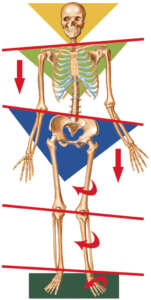 If one foot’s arch is affected due to overpronation, as seen in the picture, then the height of one lower extremity will be less than the other. The pronated foot also causes internal rotation of the lower leg. This will result in pelvis rotation along with pelvis tilt or depression. As a consequence, this will cause spine to curve in the frontal plane to bring the head to a level position, which is necessary for vision and proprioreceptive balance in the inner ear.
Excessive pronation can adversely affect gait, particularly muscle activation pattern. Recent study using electromyography found that pronated foot group when walking exhibit an increased activity of tibialis anterior, medial gastrocnemius and gluteus medius muscle. This shows how structures above the foot have to compensate for this altered biomechanics.
When laboratory induced excessive pronation was studied a statistically significant increase in internal hip rotation and anterior pelvis tilt was identified. It was concluded that ‘interaction between the foot and pelvis occurs in a kinematic chain reaction manner’. In addition, with increased bilateral foot pronation, lumbar lordosis and thoracic kyphosis are increased. Such kyphotic-lordotic posture can lead to muscle imbalance possibly causing lengthening and weakness of neck flexors, upper back erector spinae and external obliques. Moreover, it can cause neck extensor, pectoral and hip flexor shortness. If left untreated the above example of ‘imbalance between muscles that oppose each other can adversely affects posture of the body parts below and above’.
Plantar fasciitis/heel pain
Plantar fasciitis is a common complaint of many patients who suffer from chronic heel and/or foot pain. Over time, as a result of repetitive traction stress, numerous microtears occur in the plantar fascia and also on the calcaneal periosteum leading to pain and dysfunction.
The heel fat pad is designed to absorb shock during heel strike when walking or running. When efficient heel strike is not possible due to plantar fasciitis and heel pain, gait cycle becomes altered affecting ankle joint complex along with structures above.
Heel pain leads to ankle plantar flexor and toe flexor muscle impaired performance and can adversely affect windlass mechanism. Research has also shown that people with plantar fasciitis and heel pain demonstrate reduced heel loading and modified forefoot loading consistent with a strategy to offload the painful heel. Such forefoot loading requires a modified movement pattern to compensate for this dysfunction. An example would be a prolonged knee flexion of the affected leg during stance and gait with increased muscle force and stress of gastrocnemius, soleus, anterior and posterior thigh, altered depression and tilt of the pelvis leading to muscle imbalance and increased deviation of the spine above. Therefore, prolonged forefoot loading not only does overload the joints but can also cause secondary abnormal muscle activation pattern both in the lower and upper body.
The above example reflects how plantar fasciitis and heel pain can have an impact on the whole body resulting with excessive stress and pain above the foot and ankle joint complex.
Treatment
Treatment Approaches – excessive foot pronation
A chiropractor might use foot and ankle joint mobilisations, exercises and taping that control excessive pronation. Chiropractic care should be focused on restoring optimal foot and ankle joint mobility, muscle function, and normalising biomechanical control.
If excessive foot pronation is the primary cause of the problem, then management plan would focus on mobilisation, taping, exercises and foot orthotics. It is essential to achieve optimal joint mobility in excessively pronated foot along with ankle and lumbo-pelvis-hip complex if previously affected.
Exercise plan should incorporate all muscles involved with controlling pronation and facilitating the windlass mechanism along with proximal knee and hip muscles.
Due to this complex condition having much more serious effects on the whole body, involving altered pelvis, spine and head position management plan should include chiropractor cooperation with a foot specialist. This is because excessive foot pronation requires an interdisciplinary approach to provide the best possible treatment plan. For example, combining insoles with exercises has previously shown significant pain reduction. However, if foot pronation is a secondary problem caused by genu varum or hip problem, then a different management plan would need to be considered.
Treatment Approaches – plantar fasciits
A chiropractor might use manual therapy because evidence exist to support manipulation of the foot and ankle, mobilisation of the plantar fascia and lower extremity, along with soleus and gastrocnemius stretching to provide short term pain relief (1 weeks to 4 months). Heel pads can also be prescribed by chiropractor to increase benefits of stretching.
To relieve symptoms and dysfunction caused by plantar fasciitis, chiropractors can utilise kinesiology tape techniques, particularly low dye and calcaneal taping, and gastrocnemius taping for a short term (1 week) pain reduction.
In addition, active approach can be used including stretching and strengthening exercises and movement training for muscles that control foot and ankle joint to improve individual’s ability to attenuate and absorb weight-bearing forces.
A chiropractor could develop a plan that consists of soft-tissue mobilisation, passive muscle stretching and taping. These have been selected in light of the effects the plantar fasciitis has on the whole body, particularly on walking, due to pain, and muscle imbalance. Therefore, soft-tissue mobilisation is essential to maintain optimal range of motion in the affected limb, which from my own professional experience, is highly effective. Passive muscle stretching is also essential to provide pain relief, therefore improved gait, and it maintains optimal muscles length. Taping would provide better support along with pain relief. Finally, an exercise plan would be send to the patient, but ultimately the focus would be on manual treatment.